Mitral Stenosis
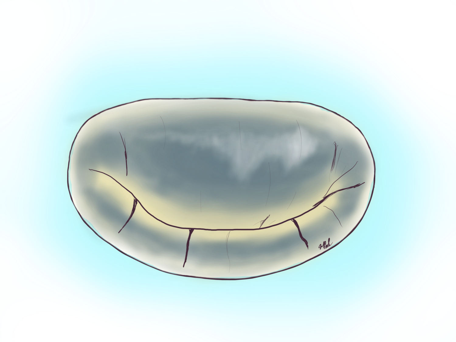
The most common reason for a tight mitral valve is Rheumatic heart disease. This problem can arise in persons who have had untreated strep infections during childhood.
Mitral STenosis
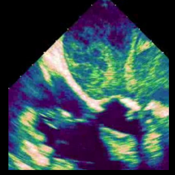
This echocardiogram pictures shows top right the blood stagnated inside the left atrium causing a pattern of swirls and the two flaps of the valve mid right screen that barely open.
A rheumatic mitral valve specimen showing heavy thickening of the valve with calcification. This valve was removed during a minimally invasive mitral valve replacement.
Rheumatic heart disease is common world-wide and has been associated with underdeveloped countries given the paucity of antibiotics to treat common throat, ear and sinus infections by Streptococcus. Surprisingly we still see and treat rheumatic heart disease in the 21st Century. The most common presentation of rheumatic heart disease is a tight mitral valve. Historically a tight mitral valve was one the original heart problems treated with surgery and became a catalyst to the advancement of the heart surgery subspecialty.
When a mitral valve is too tight the blood stagnates in the top left chamber of the heart or left atrium and pressure builds inside the vasculature of the lungs leading to pulmonary hypertension (high pressure of the lungs). This phenomenon may also lead to abnormal rhythm of the heart called atrial fibrillation. The process is progressive and leads to fatigue, shortness of breath and fluid buildup in the lungs.
A picture of a specimen rheumatic mitral valve with severe tightening . This valve was affected by rheumatic disease and one can see that should have been thin chords hold the flaps are thickened like “spaghetti” the tissue of the flap or leaflet is thickened too. The operation to replace this mitral valve was performed minimally invasive with a 2 inch incision on the right chest.
Severe mitral valve stenosis is initially treated with a percutaneous procedure performed by interventional cardiologists called catheter valve valvuloplasty, but this procedure is not for every patient. For those who are not candidate for valvuloplasty (or balloon expansion of the valve) then surgery is the best option and mitral valve replacement is the most performed operation for mitral valve stenosis.
Take Home points:
Mitral stenosis or tightening of the valve is often caused by Rheumatic mitral valve disease.
Mitral valve replacement is the most common treatment for mitral stenosis after percutaneous repairs fail.
All mitral valve replacements can be performed with a minimally invasive approach.
A transesophageal echocardiogram showing mitral stenosis
Mitral stenosis and leakage on echocardiogram
Iatrogenic mitral valve stenosis


