Other Reasons for Mitral Leakage
Ischemic Mitral Regurgitation
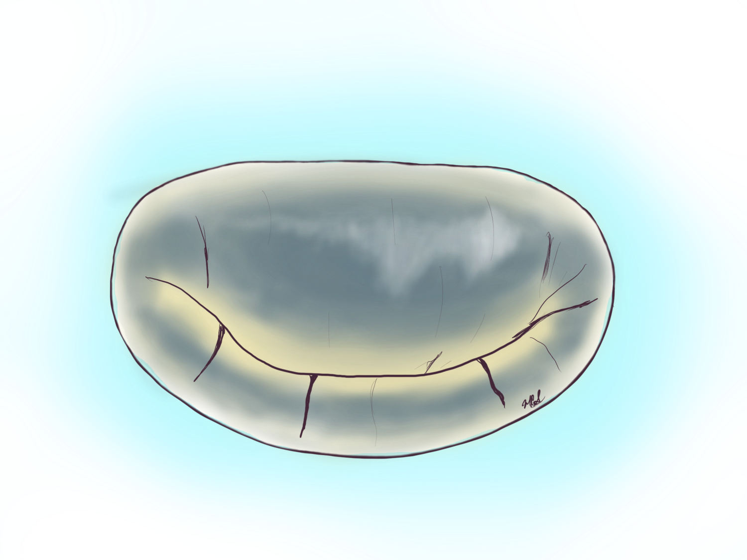
In ischemic mitral regurgitation or leakage from damage after a heart attack the lower left part of the heart or left ventricle is damaged and then scars causing retraction or pulling of the chords that hold the posterior flap (seen to the right of the image with arrow pointing to the direction of pull).
Mitral valves may leak in the absence of ruptured or stretched-out chords. Perhaps the second most common form of regurgitation is secondary to ischemic heart disease or coronary blockages.
The event we commonly know as a ‘heart attack’ is the culmination of a blockage of a coronary artery that provides oxygen-rich blood to the heart muscle. This blockage may lead to muscle death and scar formation that over time will lead to a change in the architecture/shape of the left ventricle.
Remember that the muscle of the left ventricle is intimately attached to the papillary muscle and that as such a heart attack can lead to the scaring of the papillary muscles themselves. This scar will in turn pull or restrict the chords that hold the valve. In doing so, the leaflet (usually the posterior one) is pulled down into the ventricle, with this losing the ability to prevent leakage (loss of coaptation—see image below). This form of mitral regurgitation is termed ischemic mitral regurgitation, and the medical evidence for repair versus replacement is still controversial. In these circumstances performing a replacement of the valve affords the absence of regurgitation at the cost of a prosthetic valve that, depending on the age of the patient, can have a lifespan from 7 to 20 years. The alternative is a mechanical prosthesis that will last longer, but will require the patient to be on warfarin or blood thinners for life. If a repair is performed for ischemic mitral regurgitation there is some evidence that approximately up to 20% of persons may return within a year with moderate leakage again. Making the distinction of who will leak after a repair in these circumstances is possible, but difficult.
Inside the ischemic mitral valve heart
An inside view of the heart after a heart attack. The area in tan-grey represents the area of the heart attack. The muscle inside affected includes the papillary muscle which holds the chords for the mitral valve. If the heart attack is large enough and goes untreated, the muscle can rupture and lead to fulminant mitral leakage, a life threatening condition. If the infarction is not that severe the muscle, in time, will scar and retract, pulling on the chords and flap of the valve. This is called ischemic mitral valve regurgitation or leakage.
Take Home Points:
Heart attacks can, and often cause mitral leakage.
Management of ischemic mitral leakage is still controversial with some supporting repair, and others replacement.
Surgery for ischemic mitral leakage can be performed minimally invasive.
Papillary muscle rupture from ischemic mitral valve disease
This is a picture of the muscle that holds the chords belonging to the mitral valve including the flap of the mitral valve itself. A massive heart attack led to the destruction of the muscle which later one ruptured. This is the most extreme form of what we call “ischemic mitral regurgitation” or leakage caused directly by coronary blockages.
Another view of how the muscle in the heart can die after a heart attack and lead to mitral leakage.
Heart Attack of the tip of the heart that leads to Mitral Leakage
Another example of ischemic mitral leakage. This problem originates with a heart attack, in this case at the tip of the heart which leads to scarring and ballooning of the tip of the heart or apex of the heart. This will in turn pull outwards the papillary muscles (two finger-like muscles inside the heart that hold the chords of the mitral valve) and this will splay open the valve causing leakage.
Ischemic mitral valve on 3D trans esophageal echocardiogram
This 3D transesophageal echocardiogram shows the posterior or back flap (leaflet) of the mitral valve tethered away from the anterior flap leading to a gap between the two flaps and then mitral leakage. This is phenomenon is secondary to coronary artery blockages that led to heart attacks and scaring of the muscle of the heart.


